KURSHAN LAB AT ALBERT EINSTEIN COLLEGE OF MEDICINE
WELCOME.
We use the nematode C. elegans to investigate how synapses are built. The 100 billion neurons in our brains make 100 trillion connections, or synapses, with each other. How these synapses are built is an enduring question in developmental neurobiology. The round worm C. elegans has exactly 302 neurons that make ~5000 synapses, using highly conserved molecules and principles, allowing us to use this model system to learn about the mechanisms underlying how our own synapses are formed.

Why use C. elegans?
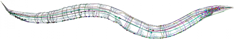
Fully mapped connectome
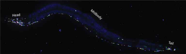
NeuroPAL (neuronal polychromatic atlas of landmarks, Yemini, et al., 2021) is a tool for identifying neurons in C. elegans
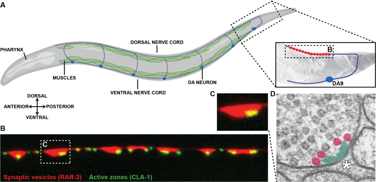
Cell-specific synaptic markers allow for high resolution imaging of individual synapses in C. elegans. A. Worm schematic showing the dorsal and ventral body wall muscles and the neurons that innervate them. B. Schematic of the DA9 motor neuron in the tail of the worm. This neuron forms a stereotyped number of synapses in a specific location onto the dorsal tail muscles. C. Confocal image of DA9 synapses in vivo expressing fluorescent synaptic vesicle proteins in red and active zone proteins in green. D. Electron micrograph of a cross section of a single presynaptic bouton (diagramed in C with a dashed box) showing the dense projection pseudocolored in green (corresponding to the active zone puncta in C).

One hour timelapse of an axon growing anteriorly towards the vulva during the L4 larval stage.